

ULTRASOUND
Head to Toe Ultrasound services:
- Thyroid Ultrasound - Scan of the thyroid gland in the neck
- Abdominal Ultrasound - Scan of the abdominal organs
- Pelvic Ultrasound - Scan of the pelvic area and organs
- Transvaginal Ultrasound - Internal scan of the pelvic organs and reproductive system in women
- Transrectal Ultrasound - Internal scan of the colon and rectum
- Obstetric Ultrasound Imaging - Scan of the foetus in pregnant women
- Liver Ultrasound - Scan of the liver
- Renal Ultrasound - Scan of the kidney and renal system
- Vascular Ultrasound - Scan of the veins and arteries

ULTRASOUND
Head to Toe Ultrasound services:
- Thyroid Ultrasound - Scan of the thyroid gland in the neck
- Abdominal Ultrasound - Scan of the abdominal organs
- Pelvic Ultrasound - Scan of the pelvic area and organs
- Transvaginal Ultrasound - Internal scan of the pelvic organs and reproductive system in women
- Transrectal Ultrasound - Internal scan of the colon and rectum
- Obstetric Ultrasound Imaging - Scan of the foetus in pregnant women
- Liver Ultrasound - Scan of the liver
- Renal Ultrasound - Scan of the kidney and renal system
- Vascular Ultrasound - Scan of the veins and arteries
An ultrasound or sonography is an imaging test that utilises soundwaves to produce pictures of internal structures. Unlike other diagnostic tests, ultrasound doesn’t involve the use of radiation but instead creates images through a series of echoes.
Typically, an ultrasound is used to examine the foetus in the mother’s womb but is also useful as a guide for biopsy and to examine the heart, liver, kidneys and stomach. The procedure helps distinguish a benign lump from a malignant tumour, evaluates blood flow and helps diagnose breast lumps, gallbladder disease, thyroid problems as well as prostate issues.
WHAT DOES AN ULTRASOUND ENTAIL?
The sonographer places a small device on the patient’s skin. Soundwaves travel throughout the body and bounce back against dense surfaces to create a picture. Soundwaves that travel at high frequencies are easily absorbed by tissues to produce high-quality images. Lower sound frequencies, however, penetrate deep layers of skin but produce poor quality images. Although a transducer is an external device, it can work within the body as well.
During the more invasive process, the radiologist attaches the device to a probe that’s carefully inserted through the oesophagus, rectum or vagina. There is slight discomfort, but generally, an ultrasound is a painless diagnostic procedure.
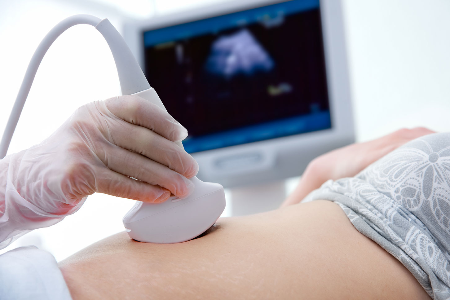
The radiologist coats the skin with a water-based gel that prevents air pockets which affect the flow of soundwaves. As the radiologist moves the transducer across the patient's skin, it captures images of the body. The visual data is sent to a computer to produce these pictures.
WHEN WILL THE RESULTS FROM THE ULTRASOUND BE AVAILABLE?
When the exam is complete, the radiologist will interpret these images and send the results to the patient’s physician. At a follow-up consultation, Dr Padi discusses his findings and treatment options with his patient and their family at his practice.
An ultrasound or sonography is an imaging test that utilises soundwaves to produce pictures of internal structures. Unlike other diagnostic tests, ultrasound doesn’t involve the use of radiation but instead creates images through a series of echoes.
Typically, an ultrasound is used to examine the foetus in the mother’s womb but is also useful as a guide for biopsy and to examine the heart, liver, kidneys and stomach. The procedure helps distinguish a benign lump from a malignant tumour, evaluates blood flow and helps diagnose breast lumps, gallbladder disease, thyroid problems as well as prostate issues.
WHAT DOES AN ULTRASOUND ENTAIL?
The sonographer places a small device on the patient’s skin. Soundwaves travel throughout the body and bounce back against dense surfaces to create a picture. Soundwaves that travel at high frequencies are easily absorbed by tissues to produce high-quality images. Lower sound frequencies, however, penetrate deep layers of skin but produce poor quality images. Although a transducer is an external device, it can work within the body as well.
During the more invasive process, the radiologist attaches the device to a probe that’s carefully inserted through the oesophagus, rectum or vagina. There is slight discomfort, but generally, an ultrasound is a painless diagnostic procedure.
The radiologist coats the skin with a water-based gel that prevents air pockets which affect the flow of soundwaves. As the radiologist moves the transducer across the patient's skin, it captures images of the body. The visual data is sent to a computer to produce these pictures.
WHEN WILL THE RESULTS FROM THE ULTRASOUND BE AVAILABLE?
When the exam is complete, the radiologist will interpret these images and send the results to the patient’s physician. At a follow-up consultation, Dr Padi discusses his findings and treatment options with his patient and their family at his practice.

Our deepest fear is not that we are inadequate. Our deepest fear is that we are powerful beyond measure
-Marianne Williamson